
Testbourne Ltd
SuperVac® Materials Division:
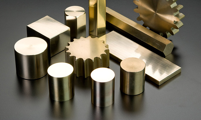
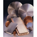
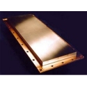
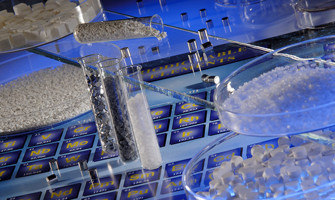
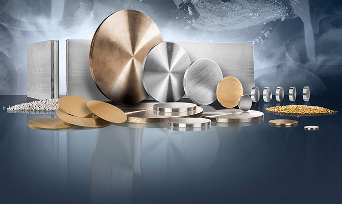
Testbourne Ltd has been supplying high purity metals, alloys & compounds to industries such as Semiconductors, Thin-films, Electronics, Electro-optics and research establishments for over 30 years. Testbourne is committed to provide the best possible service and technical expertise across a wide market area to be your first, if not single choice for an extensive selection of Metals, Alloys, Intermetallics, Compounds and Ceramics available in fabricated forms including sputtering targets, evaporation materials, powders, wire, rods & sheets. We also accommodate any custom requirements you may have.
We are proud to announce a new range of Rotatable Sputtering targets for Photovoltaic and Glass Industry.
Instrument Division:
Testbourne Limited is the representative for some of the world's leading scientific instrument manufacturers that include monitors, controllers and sensor crystals for QCM and Thin Film Technology, Sample Preparation Equipment, Thermal and E-beam Evaporation Sources, Microwave and Radio Frequency Systems, UHV feedthroughs, connectors, coaxials and viewports.
-
Superhalogens Could Yield Less-Toxic Lithium-Ion Batteries
12 December 2018Lithium-ion batteries are everywhere. They're in our electronics, our smart cars, and our power tools. As their popularity grows, so do concerns over their environmental impact. Many lithium-ion batteries contain toxic chemicals, such as fluorine, making their disposal and storage a costly issue, both environmentally and fiscally. A new study published in Angewandte Chemie International Editionpoints to at least one way the toxicity of lithium-ion batteries might be decreased. Utilizing first principles' theory, the report suggests that by altering the make-up of the batteries' electrolytes, toxic halogens can be replaced by far more environmentally friendly chemicals.
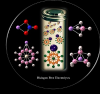
A lithium-ion battery is primarily comprised of three parts: the electrodes-cathode and anode-and the electrolyte, usually a liquid medium. As the name might suggest, the primary component of the electrolyte is lithium salt-which cycles between the negative and positive electrodes-carrying charge.Currently, among the most popular electrolytes are LiAsF6, LiBF4, LiPF6, LiClO4, LiN-(SO2F)2, and LiN(SO2CF3)2, all of which contain a halogen, says Puru Jena, a physicist at Virginia Commonwealth University and the lead author of the study. The halogen family consists of elements, including fluorine (F) and chlorine (Cl), which are toxic to humans to varying degrees. Jena and his team found that all the negative ion components of the salts belong to a special class of molecules called superhalogens, predicted by Gennady Gutsev and Alexander Boldyrev in the early 1980s. These molecules form when a metal atom at the center is surrounded by halogen atoms.
"Superhalogens are molecules that behave like halogens chemically, but they are much more electro-negative than any halogen atom," says Jena.
It should be noted that the term superhalogen is a slight misnomer: there can be halogen-free superhalogens, such as CB11H12, which still act like a halogen with high electron affinity. Jena hoped to create new superhalogens for the batteries, ones that wouldn't have the toxic elements.
To understand the fundamental properties of the current halogen-containing electrolytes, Jena and his team created models of all of the popular electrolytes. The models confirmed his suspicions: in fact, the halogens formed superhalogen constructs, which with an extra electron, form closed shells. They found that the binding of the Li+ ion in these electrolytes are smaller than that in normal salts such as LiF, making it easier for the Li ion to be stripped away and shuttled between the electrodes.
However, there was still the matter of toxicity. Jena points out that in order to remove halogens from the batteries, something equally as efficient at lithium ion transport would have to take its place. He and his team had, in the past, created molecules that acted like superhalogens but which were halogen-free. These molecules, including halogen-free CB11H12, could potentially be replacements for halogens-but only if they proved as efficient as current electrolytes in transporting the lithium.
"Through modeling, we found, actually, that our molecules were as good, if not better, than current superhalogens," says Jena. In fact, in the halogen-free molecule LiCB11H12, the lithium cation is not only bound less strongly than in the current electrolytes, but also it binds to water less strongly, another important aspect of battery production. A low affinity to water would correlate to longer battery life.
"I believe this work may have a profound effect on the development of new lithium batteries," says Boldyrev, now a chemist at Utah State University and is not affiliated with the current study.
As a next step, Jena hopes that experimentalists will synthesize some of the halogen-free lithium salts. Assuming a straight-forward synthesis, the salts can then be turned into electrolytes and run through the gamut of tests that any battery would have to perform. If successful, Jena hopes these halogen-free superhalogens might be able to replace their toxic counterparts in the near future.
By Meg Marquardt
-
Microfluidics Assembled in a Snap
12 December 2018When electricity was discovered, scientists recognized a similarity between fluid in a pipe and electrons in a wire. Wire showed resistance to flow just as pipes do. Voltage in an electrical system was analogous to pressure in a fluidic system. Current was analogous to flow rate. Now, researchers at the University of Southern California, led by engineering professor Noah Malmstadt, are reversing the metaphor to standardize microfluidic design. They report their research in a recent issue of the Proceedings of the National Academy.
"We were inspired by the idea of standardization that you see with electrical parts," Malmstadt says. "We wanted to build a common platform for fluidic circuits, just like there is for electrical circuits."
Microfluidic devices are typically made by lithography. In a typical procedure, the channel design is etched into a silicon wafer, then polydimethylsiloxane (PDMS) is poured over the wafer. After hardening, the PDMS peels off with the imprinted design. A glass or plastic substrate can be adhered to the imprinted side of the PDMS to make waterproof channels.
Lithography often requires work in a clean room. However, as Malmstadt says, "Clean room work can be pretty miserable." In order to avoid the cost (and misery) of clean room work, the modules were fabricated by high-resolution stereolithography, a type of three-dimensional (3D) printing. After designing each module, the group sent the CAD specifications to an external company that did the fabrication. Though the team assembled complex designs with their modules, assembly never took longer than an hour and everything was done on the benchtop. The team never once stepped in a clean room. Amazingly, the devices worked the first time.
"In order for a device to work properly, all the different functional domains have to be just perfect, which is pretty rare," says Michael Roper, a professor at Florida State University, who fabricates microfluidic devices in his research but was not involved in this work. "If one small channel fails or one part of the device fails, your entire device is a no-go. It's pretty rare not to have to go back and do a redesign."
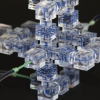
Malmstadt divided common microfluidic components into separate cubic modules with different design elements. He and his co-workers made junctions, splitters, and mixers along with unique elements such as input ports that split the flow for parallel applications and three-dimensional helices that mix two fluid streams. The cubes were standardized with 1 cm sides, 600 micrometer flow channels, and connection ports that jut out from the cube faces. Each port fits snuggly with another for snappy assembly.
To demonstrate their design philosophy, Malmstadt's team assembled a variety of devices. Emulsification modules were attached to a mixing circuit to form a range of droplet sizes at different rates. Optical sensors were added to the fabricated pieces to measure the size of the droplets while still inside of the device. The researchers coated the pre-fabricated channels with a fluoropolymer using initiated chemical vapor deposition to change the hydrophobicity of the inner walls. In their most impressive iteration, they assembled parallel three-dimensional fluidic channels for high-throughput applications, not possible with standard microfluidic designs which are almost exclusively planar.
Some systems were permanently sealed with epoxy to enhance their long-term stability, while others were disassembled and reused. Prepping the devices for reuse was simple. They ran water through the connected system, disassembled the blocks, and soaked the separated parts in alcohol. So long as the modules weren't damaged during disassembly, the reconnected joints didn't leak.
Malmstadt hopes his standardization and open-source library will make microfluidics a more prevalent research technique. "We're excited about the idea of an open design philosophy and enabling people who otherwise would be intimidated by working in a clean room to apply some of these simple design tools to their own microfluidic applications."
by Jenna Bilbrey
-
Soft, Shape-Shifting Materials Address Biological Complexity from a Bottom-Up Approach
12 December 2018Biologists tend to analyze the world by using a top-down approach, while physicists prefer to tackle problems from a bottom-up stance. When those two fields meet, however, new insights and discoveries can result. An article published in Science reports just such a breakthrough: the first soft, shape-shifting vesicle ever known to be synthesized in a lab.
"The paper is very beautiful," says David Nelson, a physicist at Harvard University who was not involved in the study. "We don't know exactly where this is headed, but it is quite remarkable and to the best of my knowledge has never been seen before."
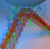
Andreas Bausch, a biophysicist at the Technische Universität München, and his colleagues built a fluid-filled pocket composed of a lipid bilayer and polymers that could actively exert forces on its surroundings. Prior to this new work, researchers could synthesize either some movable parts that mimicked the cellular skeleton, or else some parts of the vesicle. But creating an encapsulated cell-like compartment that included both of those components-and getting those components to move on their own-eluded past efforts.
To accomplish this new breakthrough, Bausch and his colleagues subjected a lipid monolayer to a special emulsion technique invented by co-author Etienne Loiseau. Then, they coupled the monolayer with a second lipid interface, paying close attention to force and time. This created the vesicle. They added microtubules-which behave like nomadic liquid crystals in the vesicle wall-to form the cytoskeleton by using a technique co-author Zvonimir Dogic developed several years ago. Finally, they applied gentle centrifugal forces to seal those two components together.
ATP-driven kinesin motors inside the vesicle, they observed, caused it to deform into various contortions as the microtubules were pushed in different directions, trying to align with one another while also being repulsed by four defects that swirled around the vesicle like comets. The vesicle's defects caused it to oscillate between a flattened and tetrahedral shape, and at times it released whisker-like protuberances. The team could tune the timing of these movements by changing the size of the vesicle or the type of kinesin motors used. "It's not dividing or crawling yet, but it is deforming into very unusual shapes," Bausch says. "When we first saw that, we were stunned."
When the researchers quantitatively analyzed the vesicles' movements and created three-dimensional reconstructions, they realized that what originally seemed like random deformations actually perfectly obeyed the laws of physics that describe the topology of nematic crystals. "Suddenly, everything made sense," Bausch says. "We recognized that everything moved by these laws."
"This work is truly important for two reasons," says Vincenzo Vitelli, a theoretical physicist at Leiden University who was not involved in the study. "The first has to do with engineering a novel class of synthetic materials made out of active components whose mechanical properties and assembly are controlled, and the second is that these synthetic structures are close enough to living organisms as to provide insights into the behavior of early life forms that marked the cross-over from inanimate to living matter."
In the very long-term, Bausch and his colleagues aim to attempt to rebuild cellular functions like cell migration and division. "The big goal is to produce something that mimics the cellular system," Bausch says. "We plan to see how far we can get by rebuilding the cell from the bottom-up."
By Rachel Nuwer
-
Mussels Inspire New Strong Underwater Adhesives
12 December 2018Strong adhesives are important for a range of technological and biomedical applications that involve high-moisture settings, such as to help repair ship hulls or heal surgical wounds. Researchers from the Massachusetts Institute of Technology (MIT) have now taken cues from nature and have designed a waterproof glue that utilizes sticky proteins from mussels and bacterial proteins found in biofilms (slimy films of bacteria that adhere to surfaces). The new material's adhesive energies are 1.5 times greater than the strongest bio-inspired, protein-based underwater adhesives reported in the literature to date, the researchers say.
"The article is very interesting and the investigators did an exhaustive amount of work," says Bruce Lee, a biomedical engineer at Michigan Technological University, who wasn't involved in the study, published recently in the journal Nature Nanotechnology. "They prepared self-assembled adhesive fibers using a 'Bottom-Up' approach, which is typically how nature constructs materials and functional tissues. It may pave a way to develop hierarchical and complex adhesive structures with improved functions."
To stick to surfaces, mussels have "feet" called byssus, which are actually strong, silky filaments. So-called mussel foot proteins (Mfps) of the byssus contain the amino acid 3,4-dihydroxyphenylalanine (DOPA). "DOPA has a specific chemical structure that allows it to stick to surfaces well," says Timothy Lu of the Synthetic Biology Group at MIT and senior author of the new study. In previous work, other researchers successfully created DOPA-based adhesives, but these glues lacked the full complexity of natural mussel adhesives, which are made up of multiple foot proteins. "Many of the existing glues have been made using single proteins on their own, or the researchers took one aspect of those proteins and mimicked it with a synthetic polymer," Lu says.
To make their bio-inspired adhesives, Lu and his team focused on the DOPA-based Mfp3 and Mfp5, which originate from the Mediterranean mussel (Mytilus galloprovincialis) and have been well characterized in the literature. They also looked at CsgA proteins, a major subunit of E. coli amyloid curli fibers-fibrous, self-assembling proteins that are necessary for the development of a biofilm and its adhesion to surfaces. Using a process called isothermal one-step Gibson DNA assembly, the researchers created two genetic fusion constructs, CsgA-Mfp3 and Mfp5-CsgA, which are gene sequences of hybrid proteins known as chimeric proteins.
The team sent their constructs to an independent company called Genewiz, which sequenced the DNA for them. E. colicells took up the genetic information, and produced the chimeric CsgA-Mfp3 and Mfp5-CsgA proteins. The researchers cracked open the bacterial cells, and then purified and incubated the fusion proteins, allowing them to self-assemble into fibrous meshes or adhesive films-simulations suggest the sticky fibers are made up of amyloid CsgA cores with Mfp domains displayed on the surface. In some cases, the team allowed the two fusion proteins to assemble together into a CsgA-Mfp3 and Mfp5-CsgA copolymer, which they hypothesized could benefit from the adhesive properties of both mussel proteins.
Lu and his colleagues tested the underwater strength of their adhesives using atomic force microscopy (AFM). They chose three AFM tips: silica, gold, and polystyrene, which represent inorganic, metal, and polymeric surfaces. The new adhesives outperformed previous protein-based underwater adhesives, as well as Mfps or curli fibers taken on their own. And at pH greater than or equal to 7.0, the adhesives exhibited better tolerance to auto-oxidation-in which DOPA oxidizes under basic conditions, decreasing adhesion energies-than Mfps alone. Additionally, the copolymers, especially those with an equal ratio of CsgA-Mfp3 and Mfp5-CsgA, were stronger than the two monomers alone.
"Using the tools of synthetic biotechnology, we've been able to combine adhesives that come from mussels and adhesive fibers from E. coli to make a strong, self-assembling adhesive that works well underwater," Lu says.
"By merging the interfacial DOPA-containing adhesive proteins of mussels with amyloid-type structures similar to barnacle cement, [Lu's team] created a hybrid system that lends itself well to fine tuning," says Herbert Waite, a biochemist at the University of California, Santa Barbara, who wasn't involved in the research. In mussel adhesion, DOPA is used for both adsorption and cohesion, but engineered DOPA-containing polymers are generally only good at one or the other; Lu's group, however, was able to take the onus for cohesion off DOPA by integrating the amyloid structures, Waite explains. "The result is a powerful wet adhesive with independently functioning adsorptive and cohesive moieties," he says. "The work is very imaginative, rigorous, and thorough."
Waite notes that one potential issue is the cost of producing the new adhesive, but streamlining the process could change this. Another challenge with the new technology, Lee adds, is "the unknown biocompatibility of these foreign and chimera proteins and unknown effects of the amyloid beta domain on the normal protein folding in our body." Scaling up the process to produce more adhesive could also prove challenging, he says.
Lu and his group are currently working on the latter issue. They are also looking at reproducing more of the proteins found in mussel feet. "There are lots of other proteins we can build to make the adhesive super sticky," Lu says.
By Joseph Bennington-Castro